


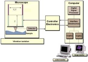
Atomic Force Microscope (AFM)
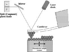 Tapping mode AFM operates by scanning a tip attached to the end of an oscillating antilever across the sample surface. The cantilever is oscillated at or near its resonance frequency with an amplitude ranging typically from 20nm to 100nm. The tip lightly "taps" on the sample surface during scanning, contacting the surface at the bottom of its swing.1 The vertical position of the scanner at each data point, in order to maintain a constant "setpoint" amplitude, is stored by the computer to form a topographic image of the sample surface.1
 Contact Mode AFM operates by scanning a tip attached to the end of a cantilever across the sample surface, while it monitors the change in cantilever deflection with a split photodiode detector.1 The tip contacts the surface through the absorbed fluid layer on the sample surface.1 A feedback loop maintains a constant deflection between the cantilever and the sample by vertically moving the scanner at each (x,y) data point to maintain a "setpoint" deflection.1 By maintaining a constant cantilever deflection, the force between the tip and the sample remains constant.1
 In non-contact mode, the cantilever is oscillated at a frequency which is slightly above it's resonance frequency, typically with an amplitude of a few nanometers (<10), in order to obtain an AC signal from the cantilever. The tip does not contact the surface, but oscillates above the absorbed fluid layer on the surface during scanning. The cantilever's resonance frequency is decreased by van der Waals forces, which extend from 1nm to 10nm above the absorbed fluid layer, and by other long range forces which extend from the surface.1
References
- Scanning Probe Microscopy Training Notebook, Digital Instruments, Veeco Metrology Group
- http://www.digitalinstruments.com/
- http://spm.aif.ncsu.edu/
