
William & Mary’s freshman phage lab goes viral for the 12th straight year
Bacteriophages — or just plain phages — are a class of virus that infect bacteria.
Phages are interesting little assemblies of protein-wrapped DNA or RNA that by most biological standards don’t even rise to the level of being alive. They’re not exactly dead, either. A phage will sit around, dormant and inert, until it bumps into a host cell. Then the phage will stab the host bacterium with its “tail,” and insert its own genetic material.
The act allows the phage to reproduce parasitically. That bacterium is never the same again. Bacteria-destroying phages have the potential to supplement — and even replace — many antibiotic drugs.
A new lab of select William & Mary freshmen takes on the study of bacteriophages each fall. It’s a program supported by the Science Education Alliance of the Howard Hughes Medical Institute called the Phage Hunters Advancing Genomic and Evolutionary Science (SEA-PHAGES) project.
“We started this in the fall of 2008 and Professor Mark Forsyth and I have been co-teaching this exciting lab for twelve years,” said Margaret Saha, Chancellor Professor of Biology at William & Mary. “We've had great success with the students and it's just been a fantastic experience, because literally on their first week on campus as freshmen, the students get to do authentic research finding these novel bacteriophages.”
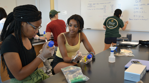
The 12th consecutive SEA-PHAGES lab at William & Mary began in earnest soon after the start of the 2019 fall semester, with 20 freshmen entering a lab in the Integrated Science Center, each bearing a bag of dirt.
Taiana James ’22, a veteran of last year’s phage lab, was getting ready for science. She had a document headed “Protocol for Making Enrichments” up on a screen. James is one of the teaching assistants for this year’s phage group. This part of the phage experience is akin to panning for gold. Instead of nuggets, the goal is to discover a new phage. And with the discovery comes naming rights.
“Last year, we only were able to isolate one phage,” she said. “And that one was mine — Herbertwm.”
Herbertwm was duly entered into the database at phagesdb.org — and James made a presentation on her discovery at the HHMI Janelia Research Campus. It also turned out that another “phagelet” called Bernie is also needed for infection: “We are still intensively investigating these novel and exciting findings,” Saha said.
James said this year’s lab will use two new bacterium hosts, hoping to isolate more phages. Forsyth added that past William & Mary phage labs worked as part of a HHMI-funded consortium of schools, all using mostly the same Mycobacterium species (Mycobacterium smegmatis).
“We probably isolated thousands of phages,” Forsyth said. “And, over time, I think we’ve largely exhausted that pool of phages. So, we’ve moved on to different Mycobacteria species to try to optimize the discovery of novel viruses — and, most importantly, novel genes that are within the viruses.”
Phage lab participants have an excellent chance of finding a novel specimen — a phage completely new to science. Phages are unbelievably numerous, both in absolute numbers and in variety. Many William & Mary students have discovered, annotated and studied novel bacteriophages. In fact, the students in the first W&M phage lab found a new phage in the muck of William & Mary’s landmark Crim Dell. They named it CrimD, and as a specimen it was interesting enough for a lab at the University of Pittsburgh to incorporate it into a study looking for organisms to combat tuberculosis.
“Bacteriophages are viruses that attack and infect bacteria — and only bacteria,” Forsyth notes. “They're perfectly safe for humans, so we're not exposing the students to any virus that could infect them.”
In fact, bacteriophages have been used as antibacterial agents for decades. The plot of the 1925 Sinclair Lewis novel Arrowsmith follows a researcher’s work to develop a phage strain to combat a bubonic plague outbreak. The development of effective antibiotic drugs sidelined interest in (and support for) phage research.
Recent revelations about “superbugs” and bacteria that are evolving resistance to antibiotics have rekindled interest in bacteriophage therapy, and Saha points out that the freshmen enrolled in the phage lab are getting in on the ground floor of what promises to be a resurgence in phage virology. She added that the William & Mary program has evolved along with the science.
“For the first six years of the program we used the host called Mycobacterium smegmatis. It's a very common bacterial species that one finds in the soil,” Saha said. “But then, we’re William & Mary, so we wanted to expand the host range. We wanted to take some risks and use different hosts in order to find novel phages.”
“There is a huge amount of clinical relevance to using a mycobacterial host,” Saha said, and discovery of a phage that infects some of the species most troublesome to human health would be, literally, just what the doctor ordered in a time of growing bacterial drug resistance.
Not all of the 190 or so mycobacterium species are dangerous to humans. Saha stressed that the lab works with bacterial hosts rated BSL1 — no real danger of human infection, therefore benign cousins of the dangerous mycobacteria species.
William & Mary’s phage lab has a couple of other distinguishing characteristics, beyond getting first-year students doing real science when they first hit the Integrated Science Center. The first is the persistence of interest in phage research. Saha and Forsyth have many phage lab alumni who come down with a serious case of the phage (research) bug.
“I have a whole group that's continuing,” she said. “They used to be part of freshman phage lab, now they're rising sophomores, juniors and seniors and are continuing their research”
The second characteristic is how far beyond the original phage lab experiments students take their projects. Phage lab alumni are now employing state-of-the-art techniques such as RNA-Seq and sophisticated bioinformatics analysis, and are extending the boundaries of phage research.
But each new phage lab begins as this one did: with students collecting material for analysis.
“Everyone have their soil?” Saha asked phage lab members as they gathered to execute the first steps in finding their bacteriophages. “Their nice, gooey, dirty soil?”
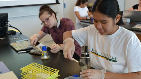
They did. Each lab member had a ziploc bag containing a few tablespoons of nice, gooey, dirty soil. Saha stressed the importance of keeping accurate records.
“What were the GPS coordinates where you got your soil — longitude and latitude?” Saha said. “Use your phones. Also: time of day? How deep did you dig? Was the soil wet? Was it near water?”
Saha explained that there are two ways to go about the task. The first, direct plating, checks for phages that might happen to be in the soil samples.
“But in all of the 12 years that we’ve been doing phage lab, we’ve never found a single phage by direct plating,” Saha said. “So we’re going to do enrichment today.”
As Saha spoke to the lab, Teaching Assistants Elizabeth Do ’21 and Hermela Zerihun ’21 were back in the prep area preparing a fresh batch of a bacteria-enriched solution known as “broth.” The soil goes into the broth, and phages infect the bacteria in the broth and multiply. It’s much easier to find a group of phages than a single specimen, as population of bacteriophages make their presence known in the form of a “plaque” or clear spot in a dish.
The TAs wheeled out a tub filled with flasks of fresh broth. A flask marked with yellow tape indicated broth rich with Mycobacterium dienhoferi; white meant M. rutilum. As the lab members began to use tongue depressors to spoon some of their soil into the flasks, Saha called out their attention to a fact that blended caution and encouragement.
“Remember: You may not find anything in your sample. That’s O.K.,” she said. “You’re doing real science. Real science. And that’s the way it works.”