
One more reason to love the striped bass: antimicrobials
UPDATE: Myriam Cotten’s paper has been selected for inclusion in a special round-up issue of the Journal of Biological Chemistry (JBC) devoted to immunometabolism.
“Immunometabolism has emerged as a very exciting area of research in immunology,” JBC editors wrote in their introduction to the special issue. “The number of papers published since 2010 has skyrocketed as immunologists dust off their old biochemistry text books and re-learn the wonders of glycolysis, the TCA cycle, the pentose phosphate pathway and the intricacies of amino acid metabolism.”
Cotten’s paper was included in a section titled “Innate Immunity.” Her work examined antimicrobial peptides that are produced naturally by striped bass, as described in the original story below.
“We looked through a large number of papers to come up with what we felt best represented the exciting advances in immunometabolism in recent years, and we are very pleased to include your paper in the group,” Jonathan Griffin wrote to Cotten. Griffin is associated with the American Society for Biochemistry and Molecular Biology, which publishes JBC.
Cotton’s work also received attention in the outside media.
It’s hard to think of a fish with a higher across-the-board value than the striped bass — or rockfish, as it’s known in the Chesapeake Bay region.
Morone saxatilis is esteemed by anglers who fish in freshwater as well as saltwater. It’s a valuable commercial species, and therefore earns a top listing in the seafood section of many a restaurant menu and fishmonger’s stall.
A team of scientists at William & Mary led by Myriam Cotten is investigating yet another virtue of the striped bass: The fish contain biomolecules that have shown promise for therapeutic use in human medicine.
Cotten, an associate professor in the university’s Department of Applied Science, is a co-author on a recently published paper, “Copper regulates the interactions of antimicrobial piscidin peptides from fish mast cells with formyl peptide receptors and heparin,” in The Journal of Biological Chemistry.
She explained that the peptides studied in the paper are varieties of the molecular weapons used by an animal’s immune system, and produced by mast cells — specialized white blood cells. In this case, the subject animal is a fish, and so they are “piscidin” peptides.
The paper notes that fish are subject to a barrage of pathogens — bacterial, viral, parasitic and fungal. Fish, of course, swim and breathe in what sometimes can be a soup of pathogens. Some 65 percent of infections get their start in biofilms and to stay healthy, fish have evolved powerful immune systems to fight off infection.
Cotten says she often works with other scientists, particularly when it comes to in vivo examinations: “I do the fundamental research — I love that! But I don’t do in vivo,” she explained. “That’s why this is collaborative work.”
For instance, she said that in 2015, after studying them for a decade, she found that her peptides could bind copper. It was an important discovery.
“Copper ions form radicals, and radicals can attack neighboring biological molecules, latching on and damaging certain chemical bonds,” Cotten explained.
She collaborated with Hao Hong at the University of Michigan, who tested her copper-charged peptides in vivo applied to a cancer tumor on a mouse. The results, while preliminary, are promising, Cotten added.
The copper-charged radicals could be an important new weapon in the wars against tumors and infections, and the mechanisms described in the J. Biol. Chem paper are the necessary next steps on the route to clinical trials. The paper describes the peptides as Swiss army knives that not only directly attack bacteria but also activate the immune cells of the host organism to help fight infections.
As she works toward better understanding of the biochemical mechanism of piscidins and other biomolecules that could one day be used to fight off infections in humans, Cotten appropriately describes herself as a biophysical chemist.
“That means I study biological systems with physical tools,” she explained. Crystallography is one of the go-to physical tools for chemists, but Cotten studies biological membranes and notes, “Everything that has to do with a membrane, that binds a membrane, that targets a membrane, is very hard to study with crystallography when it is bound to that membrane.”
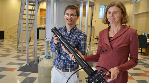
Therefore, Cotten’s own go-to tool is nuclear magnetic resonance, or NMR. She is continuing her investigation in the NMR lab in Small Hall on the William & Mary campus, working with a group that includes research scientist Dr. Alex Greenwood, an NMR specialist, and supported by funding from the National Science Foundation.
“NMR happens to be one of the best techniques to interrogate samples that do not crystallize,” Cotten said. “When you are looking at an antimicrobial substance that is very likely attacking membranes or maybe has internal targets, like DNA, there’s really no atomic-level method other than NMR that can study non-crystalline samples.”
Nuclear magnetic resonance is a sensitive and exacting technique — or rather set of techniques. Long before she came to William & Mary, Cotten was familiar with the NMR work going on here, particularly the work of Robert Vold, a former member of the faculty in the physics and applied science departments.
“When I was a graduate student, I was in awe of the work of Professor Vold, ” Cotten said. “I still have stacks of his papers that I printed 20 years ago. And I never met him until I got the job here.”
Cotten began to work in the big magnet lab at Small Hall, using the 17.6-tesla, 750 megahertz magnet that Vold used. But she could not just walk up and put her samples into the magnet. Vold, along with Gina Hoatson and a number of other users of the big magnet, had been using it to study non-biological samples. Cotten’s work involves examination of biological membranes.
“When I came here, the instrument was set up for materials. I had to buy new probes to do NMR here,” she explained.
NMR users like to compare the magnet to a drill and the probes to drill bits: A drill can use any number of bits, depending on the materials. The type of “bits” Cotten needs to study membranes are called static probes. One of her collaborators, Peter Gor’kov of the National High Magnetic Field Lab at Florida State, built the new probes for her.
The samples are oriented on glass plates and then they go into the throat of the magnet, the coil. The magnet zaps the sample with a magnetic field. The atomic nuclei in the sample have their own electronic environment and resonate at unique frequencies within the magnetic field.
The probe sits inside the magnet, about two feet, using radio waves to pick up the transfer in energy level in a signal that, once it’s decoded, can yield up a great deal of detail about the molecular structure of the sample.
Cotten says she is “deeply passionate” about using NMR because it allows for the study of the movement of molecules, not just their structure. And molecular movement is quite important to her understanding of phenomena associated with membranes.
“Molecules don’t work by being frozen in space. Molecules move,” she explained. “If you have a substance that’s antimicrobial, it needs to move to a site where it attacks a cell. When it’s there, it needs to change the structure of what it’s bound to in order to damage that site. It’s very dynamic.”
Cotten and her team are working to install the new biomolecule-friendly probes in the NMR lab to continue study of the fish peptides, which show promise for an ever-widening array of clinical applications.