
Andriy Fedorov Wins Top Award at Graduate Research Symposium
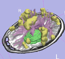 Neurosurgeons rely on MRI images to map the location of brain
tumors and guide their work in separating tumors from healthy brain
tissue. High-resolution scans taken under optimum conditions before
surgery reveal cellular differences that are not visible to the eye.
Neurosurgeons rely on MRI images to map the location of brain
tumors and guide their work in separating tumors from healthy brain
tissue. High-resolution scans taken under optimum conditions before
surgery reveal cellular differences that are not visible to the eye.
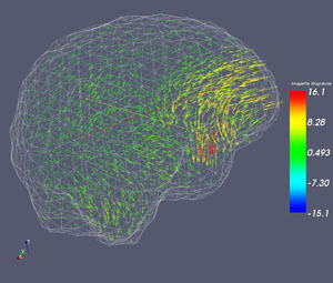
Yet the surgery itself causes the brain to change shape, shifting the tumor's location. Ideally the surgeon could view real-time scans throughout the surgery and follow the tumor's changing location precisely.
Andriy Fedorov is demonstrating the potential for just this kind of solution. A Ph.D. student of Prof. Nikos Chrisochoides, Andriy has worked for two years putting together a sequence of processes involving an algorithm that registers the pre-surgical MRI image to images taken during the surgery.
Andriy's research project is titled, Near-Real-Time Nonrigid Registration for Image Guided Neurosurgery Using Commodity and Grid Computing. At the College's sixth annual Graduate Research Symposium, held in March 2007, his project won the Award for Excellence in Scholarship in the Natural and Computational Sciences.
In Andriy's research scenario, the surgery takes place within a "double-doughnut" MRI configuration that allows scanning while still providing the surgeon access to the brain cavity. "Speed is a key factor," Andriy notes. "We have to be able to take new images during surgery, register them to the pre-surgical image, and return updated information to the surgeon as quickly as possible."
Because MRI images are digital, it's possible to compare and manipulate the data mathematically. "To register the images we applied an existing algorithm, which we tweaked to improve speed and, in the long run, its accuracy," said Andriy. "Our requirements for the full registration sequence are accuracy, robustness, and computational feasibility."
During his first year into the project, Andriy Fedorov developed a tool that constructs the tesselation of the brain from an MRI image. Such tesselation, also known as "mesh," enables biomechanical modeling of brain deformation for MRI registration. Last year, Andriy Fedorov and his fellow student Andriy Kot, worked together and successfully delivered the implementation of registration, which computes the result here at W&M and sends it to the surgeons at Brigham and Women's Hospital (BWH) in Boston in less than 5 minutes. For the first time the computation of such complexity was completed within the time constraints of the surgery. Since then, the implementation has been used routinely at BWH.
 Andriy emphasizes that this result was made possible by a
dedicated team effort. In addition to his colleagues in the Parallel
Experimental Systems Lab in the Computer Science Department, the team
includes world-recognized experts from around the globe. Medical image
processing expertise comes from Olivier Clatz from INRIA
Sophia-Antipolis, France, Computational Radiology Lab led by Simon K.
Warfield and the Surgical Planning Lab led by Ron Kikinis, at
Children's Hospital and BWH respectively. Neurosurgeons Alexandra Golby
and Peter Black at BWH, where the open MRI scanner is located, are also
contributing to the investigation. This work is part of a very large
interdisciplinary team put together during the last 14 years by Ferenc
Jolesz at Harvard Medical School.
Andriy emphasizes that this result was made possible by a
dedicated team effort. In addition to his colleagues in the Parallel
Experimental Systems Lab in the Computer Science Department, the team
includes world-recognized experts from around the globe. Medical image
processing expertise comes from Olivier Clatz from INRIA
Sophia-Antipolis, France, Computational Radiology Lab led by Simon K.
Warfield and the Surgical Planning Lab led by Ron Kikinis, at
Children's Hospital and BWH respectively. Neurosurgeons Alexandra Golby
and Peter Black at BWH, where the open MRI scanner is located, are also
contributing to the investigation. This work is part of a very large
interdisciplinary team put together during the last 14 years by Ferenc
Jolesz at Harvard Medical School.