
Equipment Descriptions - William & Mary Campus
Please contact the ARC Core Labs for questions and assistance.
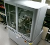
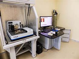
Profilm3D Surface Profiler, ISC3-1223 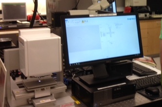
Profilm3D surface profiler provides sub-nanometer vertical resolution using vertical-scanning interferometry (VSI) and phase-shifting interferometry (PSI). Softwarecapable ilities include measurements for surface roughness, topography, and step heights. Common roughness parameters can also be measured on both flat or curved surfaces. (Specifications from Filmetrics)
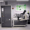
Trift II Time of Flight Secondary Ion Mass Spectrometer (ToF-SIMS), ISC3-1223
ToF-SIMS is an analytical technique that uses a primary ion beam in an ultrahigh vacuum environment to probe the surface of a solid material. ToF-SIMS provides spectroscopy for characterization of chemical composition, imaging for determining the distribution of chemical species, and depth profiling for thin film characterization. Performance capabilities include one parameter tuning used for insulating samples, a library of high mass resolution reference spectra, and high-speed SIMS imaging setup and acquisition. Analytical applications include statistical analysis of trace (ppm) contamination and multiple defects on silicon wafers and magnetism analysis of surface contamination, drug delivery cross-section media, high spatial resolution surface and cross-section samples, and failure analysis samples. (PHI Trift II ToF-SIMS Specifications)
Phenom ProX Desktop Scanning Electron Microscope (SEM) with EDS, ISC3-1223 (Specifications)
The Phenom ProX desktop SEM includes an optical color camera and an elemental identification feature with fully integrated EDS that will enhance our current analysis capabilities. Advantages of the Phenom ProX system include easy operation and simple sample preparation. A wide range of sample types, such as metals, polymers and biologicals, can be viewed at magnifications between 80x to 130,000x. Samples up to 32 mm in diameter and 100 mm in height can be loaded in less than 30 seconds. The Phenom ProX system includes a 19 inch flat panel touch screen monitor which is used to view, compare, and measure sample images at a maximum 2048 x 2048 pixel resolution. The Phenom ProX is a very compact, quiet, and a low maintenance addition to the lab. Images can also be saved to a USB flash drive for future use. (Additional Information)
Hitachi S-4700 Field Emission Scanning Electron Microscope (FE-SEM) with Energy-Dispersive X-Ray Spectroscopy, ISC2- 2107A
The Hitachi S-4700 FE-SEM is a cold field emission high resolution scanning electron microscope. The FE-SEM has a magnification range between 30x and 500,000x. Spatial resolution of up to 1.5 nm at 15 kV, 12 mm WD and 2.5 nm at 1 k, 2.5 mm WD. Digital images may be acquired in BMP, TIFF, or JPEG file formats. Elemental analysis of a sample can be performed using EDAX Energy-Dispersive X-Ray Spectroscopy capabilities. (User Instructions, Energy Dispersive X-Ray Spectroscopy, and Sample Holder Assembly Video)
Phenom G2 Pro Tabletop SEM, Small Hall Maker Space 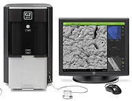
Empyrean Series 2 X-Ray Diffraction System (XRD), Small Hall, Lab 273
With the Empyrean, Panalytical has set the new standard for a multipurpose diffractometer. In developing the ultimate X-ray platform for the analysis of powders, thin films, nanomaterials and solid objects, the Panalytical R&D team has redesigned all key components of the X-ray diffractometer from the ground up. It is Panalytical's answer to the challenges of modern materials research, where the lifetime of a diffractometer is considerably longer than the horizon of any research project. This multi-purpose x-ray diffractometer can be used to analyze powders, thin films, nanomaterials and solid objects. The PreFIX concept for optics and stages allows changes in variables and applications without the need to realign the system. More information about the XRD is available from Panalytical in the User's Guide and on the Malvern Panalytical website.
Bruker Tracer III-SD Handheld XRF Spectrometer, ISC3-1223 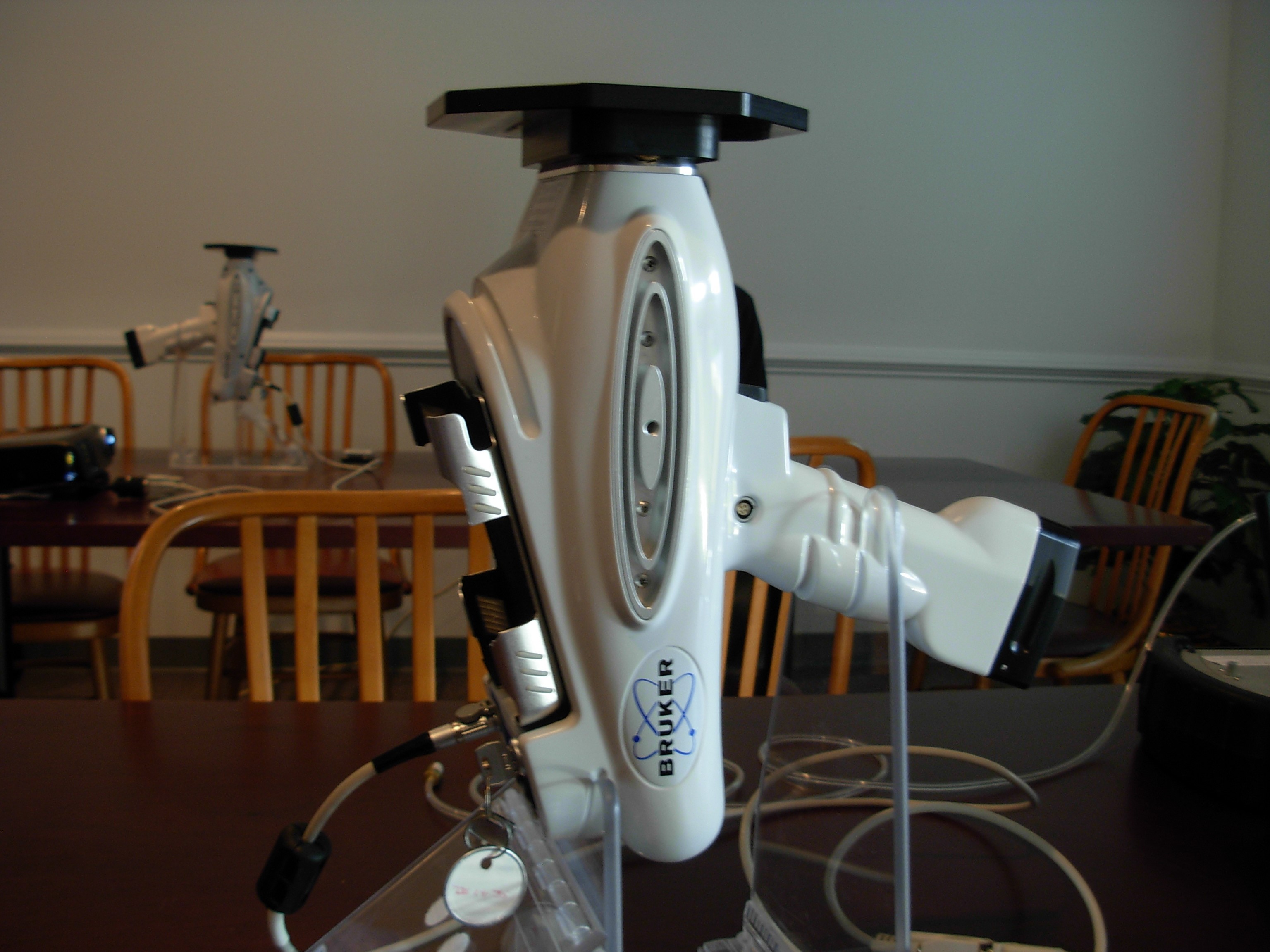
This handheld XRF allows for x-ray diffraction and elemental analysis of samples with greater flexibility over bench-top models while using the same vacuum technology. The XRF also utilizes lap-top based analytical software with real time spectral displays and spectral displays with complete peak insentification.
Technical Details: (Additional Specifications)
- XFlash Silicon Drift Detector (SDD) provides high speed data acquisition and better resolution
- Typical resolution: 154 eV at 100,000 cps
- X-ray tube: Rh target, max. voltage 40 kV
- Weight: 2 kg (4.49 lbs) with batteries
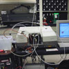
Nicolet Nexus 670 Fourier Transform-Infrared Spectrometer (FT-IR), ISC3-1223
FT-IR spectroscopy is used for molecular characterization of solids, liquids, and gases. This includes a wide range of materials which can easily be evaluated. The FT-IR features a 11,000 - 375 cm -1 range and 0.125 cm -1 resolution with Transmission, ATR and DRIFT operations available. The FT-IR also includes a DTGS detector. Currently, the FT-IR is optimized for mid-IR spectroscopy. The time needed to collect data under best resolution is 8 minutes. The accessories available are identified below:
- The Smart Orbit diamond attenuated total reflectance (ATR) accessory accommodates flat samples such as polymer films and requires little sample preparation.
- The Spectra-Tech Collector II diffuse reflectance (DRIFT) accessory provides surface analysis of powders and particles. Sample concentrations can range from parts per thousand to neat.
HIROX RH-2000 High Resolution Digital-Video Microscopy System, ISC3-1223
The Hirox RH-2000 2D / 3D digital microscope provides high resolution observation, accurate and fast measurements, fully motorized XYZ axis for 3D tiling, and a state-of-the-art complementary metal oxide semiconductor (CMOS) sensor with improved light sensitivity and very low image noise. The resolution is higher than Full HD (1920 × 1200), at a very fast 50 FPS (special 100 FPS mode). Options include Fast PC and Full HD Screen via USB3.0 connection. 3D models can easily be created with an integrated stepping motor that allows for fast and accurate scanning. 3D data can be quantified by associating the profile graph with the image display area so 3D height can be measured along with and the extract angle and radius data. With the combination of tiling and 3D modeling,the field of view can be increased to more than 170 times while retaining a high optical resolution. Point height measurements are possible in both 2D and 3D rendered images. Volume and area can also be measured on the 3D object by adjusting the horizontal cross section and clicking on the area of interest. In addition, surface roughness can be measured using the 3D software. Zoom capabilities ranging from 10x to 7000x magnification can be achieved by using a variety of lenses.
HIROX KH-7700 High Resolution Digital-Video Microscopy System, New Location TBD 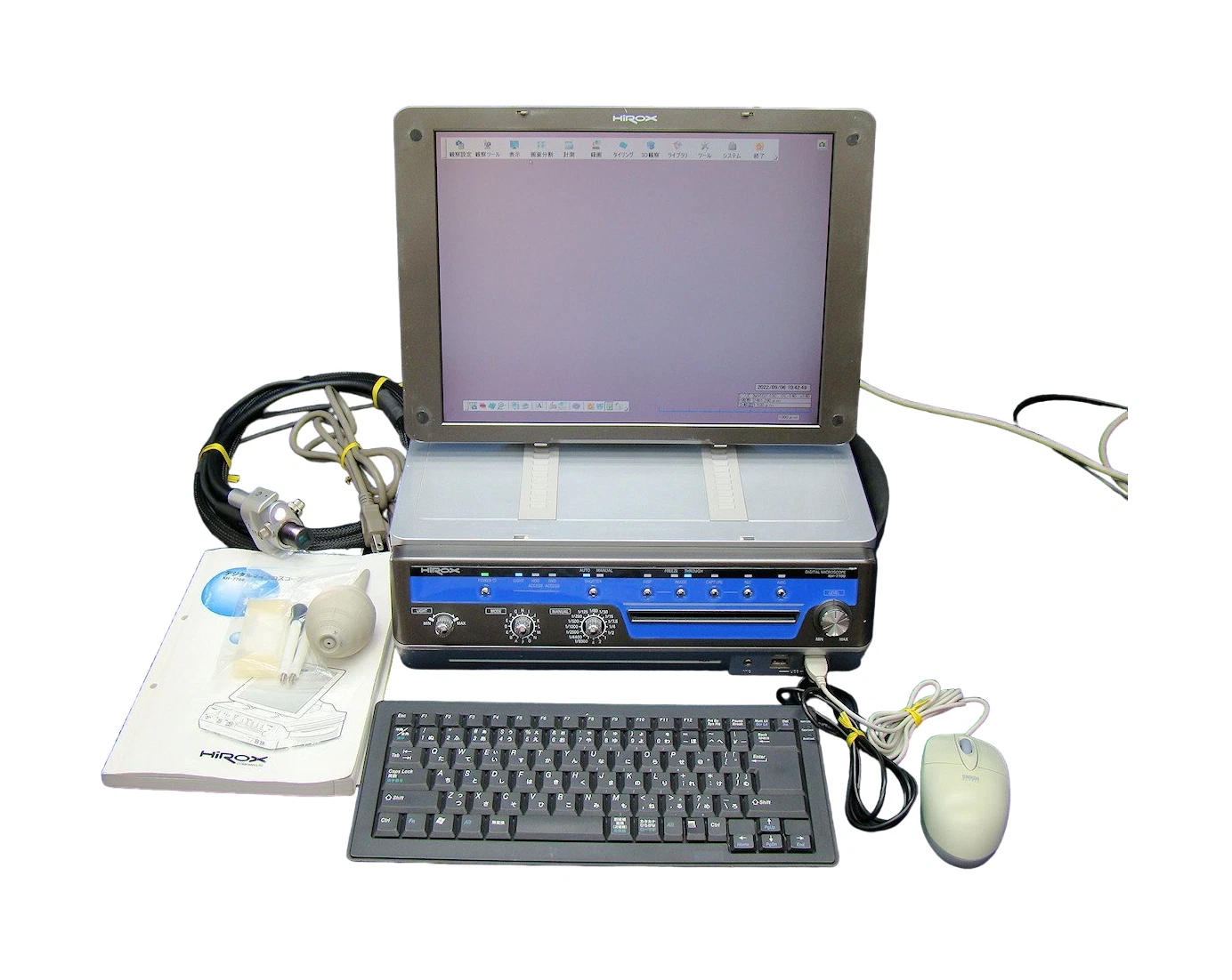
The HIROX KH-7700 digital microscope provides enhanced 3D image synthesis and image comparison. The report generation feature will save a significant amount time and standardize our documentation. This compact system includes a digital camera, light source, LCD monitor, computer and software which are all integrated into one compact unit. Zoom capabilities ranging from 10x to 7000x magnification can be achieved by using a variety of lenses.
Alpha-SE Spectroscopic Ellipsometer, ISC3-1223
Ellipsometry is a technique used to characterize optical properties and thicknesses of thin films by measuring the change in polarization state of light reflected from the surface of (or through) a sample. The alpha-SE can be easily used to measure both thin film thickness and refractive index of all materials including dielectrics, semiconductors, and organics. Measurements are completed in three simple steps. First, place the sample on the stage and then choose the model from the software that matches your film. Press the measuremnet button and your results are available in seconds.
Specifications and additional details can be found on the manufacturer's website at jawoollam.com. The video "Introduction to the alpha-SE Spectroscopic Ellipsometer" is also provided by JA Woollam.
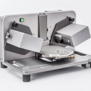
Bruker Dektak XT Surface Profiler, ISC3-1223 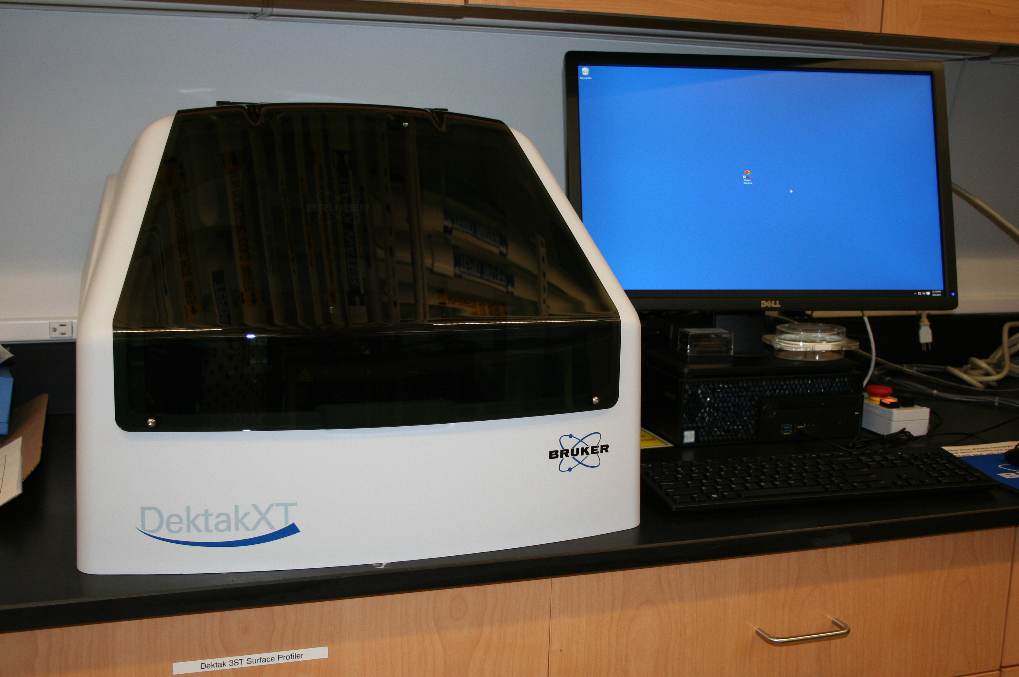
The DektakXT® stylus profiler features a new design that enables repeatability of 4Å and up to 40% improved scanning speeds. The DektakXT can perform the critical nanometer-level film, step and surface measurements needed to power future advances in the microelectronics, semiconductor, solar, high-brightness LED, medical, scientific and materials. This is the first stylus profiler to implement a single-arch design, the first to incorporate a true-color HD optical camera, and the first to use 64-bit parallel processing architecture to achieve optimal measurement and operating efficiency. Vision64, Bruker's 64-bit parallel processing operation and analysis software, enables faster loading of 3D files and faster applications of filters and multiscan database analyzes.

Dektak 3ST Surface Profiler, MS Hall, Lab 325
This is an extremely accurate high precision surface measurement system. Analytical functions include parameters for roughness, waviness, step height, and geometry. Available styli include sub-micro, 2.5 mm, 12.5 mm, and 25 mm.
Shimadzu Vickers Microhardness Tester Type M (Vickers Hardness Tester), ISC3-1223
The Vickers Microhardness Tester is primarily used for fine wires, thin plates, super-hardened surfaces, thin films, implanted materials, and the precise hardness of other materials. It is equipped with a Computer Assisted Measurement System (CAMS) and imaging capabilities.
The Vickers Microhardness Tester uses a square based diamond pyramid whose opposite sides meet at the apex at an angle of 136 degrees. The Vickers number is known as HV and is calculated using the following formula:
HV (applied load) = 1.854 (F / D 2 )
(F = applied load in Kgm-force)
(D 2 = area of indentation in mm)
MakerBot Replicator Mini Compact 3-D Printer, ISC3-1223 (2 MakerBots Available for Reservations)

The Makerbot Replicator Mini is housed in a rigid frame which is open at the front, on the sides, and on top, permitting easy access to the print bed and easy viewing of prints in progress. Printing can be initiated from a computer over a USB or Wi-Fi connection, as well as over Wi-Fi from an iOS or Android phone or tabloid with the MakerBot Mobile app installed. A camera in an upper corner of the Mini allows monitoring of print jobs from a computer or mobile device. The Mini's build area is 3.9 x 3.9 x 4.9 inches. Small (0.5 lb) spools of 1.75 mm polylactic acid (PLA) filament are required and are available at the lab in several colors.
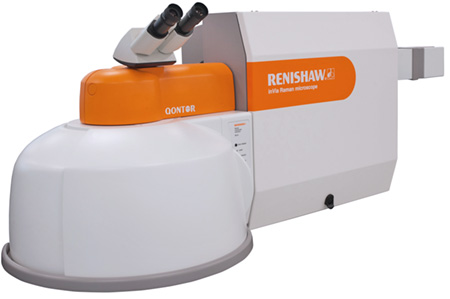
The Renishaw inVia Raman Microscope is easy to use research-grade confocal Raman microscope that delivers outstanding performance and reliable results which include acquiring detailed chemical images and highly specific Raman data from discrete points. Analysis may be performed on both large volumes and minute traces of material.
Perform all types of Raman measurements:
- Time series - How your sample alters over time
- Temperature ramps - Phase changes with a hot/cold cell
- Line scans - Profile your sample across the surface or into its depth
- Area mapping - Horizontal images at fixed focus across the topography, or vertical slices. More details…
- Volume scans - 3D views of your transparent sample's internal structure
- Transmission mapping - Analysis large volumes of material and produce depth-averaged 2D images of bulk material homogeneity
- Specialist measurements - Trigger data collection from your own equipment (control system on a synchrotron beamline)
The easy-to-use software allows you to collect the data you need, analyze and display it as you want to.
AFM Dimension Icon, Bruker, ISC3-1223
 The Dimension Icon Atomic Force Microscope (AFM) incorporates the latest evolution of Bruker's industry-leading nanoscale imaging and characterization technologies on a large sample tip-scanning AFM platform. The Icon's temperature-compensating position sensors render noise levels in the sub-angstroms range for the Z-axis, and angstroms in XY. This level of performance has established the new generation of what Atomic Force Microscopy should be.
More technical information is available in the Dimension Icon brochure from Bruker.
AFM Dimension 3100, Bruker, Jefferson Lab, TED Building, Newport News
Specifications/Capabilities:
- Contact mode, tapping mode, and phase imaging, electrostatic force microscopy, magnetic force microscopy, nanomanipulation, and nanolithography.
- Small sample sizes can be supported with a maximum wafer diameter of 200 mm and a maximum substrate thickness of 12 mm.
Buehler Minimet 1000 Polisher/Grinder, Small Hall Lab 168 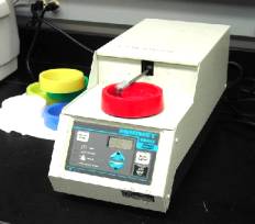
Single-specimen polishing and lapping operations include:- Fine grinding
- Rough polishing
- Final polishing
Variable load control: 0-10 lb
Variable speed control: 0-50 rpm
Instructions for Use (pdf)