


Got it on eBay…
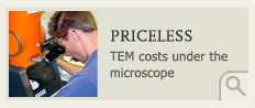
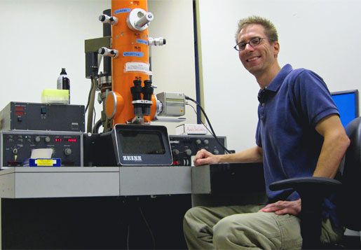 Scoping it out Virologist Kurt Williamson puts the department’s "new" transmission electron microscope through its paces. Williamson bought the lab instrument on eBay, paying a small fraction of the price of a new TEM. Students from the HHMI-supported phage lab for William & Mary freshmen will use the microscope to examine phages they’ve isolated from soil samples. Photo by Joseph McClain
…and our transmission electron microscope is running just fine, thanks
It looked like the 109 was dead, not that it owed us anything.
The 109 had lived a good, long life of service to science. Kurt Williamson first saw the 109 in 2008, when he interviewed for the assistant professor position in William & Mary’s Department of Biology. It was old, but in robust health and very much in use. The 109 is a Zeiss transmission electron microscope (TEM), an elegant arrangement of optics, plumbing, mechanics and electronics that allows biologists to see—and photograph—things as small as viruses. They function much like an old-school microscope, except instead of directing light up through the specimen, TEMs shoot a beam of electrons down a vacuum chamber and through the specimen, providing a resolution that allows scientists to examine details at the sub-cellular level.
Williamson was reassured by the presence of the Zeiss 109 in Millington Hall. In graduate school, he had done a great deal of work on a Zeiss 902, a similar model, and he knew he could begin being productive with the William & Mary instrumentation with no learning curve.
“It’s like learning to drive on a Honda Civic fancy model, then driving a Honda Civic base model,” he said. “There’s a few less bells and whistles, but it’s the same setup. I knew immediately how to use it.”
Williamson, a virologist, got the
job. He quickly became one of the three William & Mary biologists leading
the freshman bacteriophage laboratory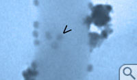 . Bacteriophages—or just plain phages—are
viruses that infect bacteria. The phage lab is an initiative sponsored by the
Science Education Alliance of the Howard Hughes Medical Institute. For
freshmen, it’s a fast track into a genuine research experience, mentored by a
trio of biology professors that, in addition to Williamson, include Margaret
Saha and Mark Forsyth.
. Bacteriophages—or just plain phages—are
viruses that infect bacteria. The phage lab is an initiative sponsored by the
Science Education Alliance of the Howard Hughes Medical Institute. For
freshmen, it’s a fast track into a genuine research experience, mentored by a
trio of biology professors that, in addition to Williamson, include Margaret
Saha and Mark Forsyth.
HHMI’s Science Education Alliance (SEA) is a national initiative to enhance science education and to increase the number of scientists produced in the United States. Saha, William & Mary’s Chancellor Professor of Biology, is the principal investigator on the HHMI grant. She has received a total of $6.2 million in funding from HHMI since 1998.
Each fall, the lab begins with a phage hunt, as the freshmen scoop up soil samples, then employ a series of laboratory techniques to isolate phage specimens. It’s not uncommon for one of the students to discover a phage that’s previously unknown to science. Such was the case in the first year of the HHMI lab, when the freshmen isolated the now-famous CrimD from the muck of Crim Dell.
Seeing is inspiring
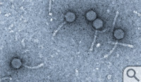
Williamson introduces the freshmen to the TEM as part of the comprehensive scientific examination of the isolated phage samples. The HHMI phage labs are designed to give college freshmen an introduction to the latest microbiological techniques for examining the viruses, up to and including the sequencing of the phage genomes. Compared to gene sequencing, TEM is a rather basic analytical technique, but Williamson says TEM has a number of virtues that make it the workhorse of microbiological labs. The TEM preparation is quick, he said, and gives the user a visual image of the sample.
“That’s important, because we can
sort out what phages we might have seen before from those we might not have,”
he said. Phages 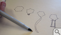 come in four body types, distinguishable quickly by the kind of
tail. There are no-tail phages, phages with a short stub of a tail and phages
with a thick, contractile tail. So far, most of the phage specimens isolated in
William & Mary’s HHMI lab have been of the Siphoviridae body type, with long, flexible tails, Williamson said.
He added that TEM reveals other distinguishing characteristics such as tail
length and head shape. TEM allows for a quick sorting of phages by morphology,
or body type, he explained, and the specimens with the more interesting
structures can be prioritized for gene sequencing. The visual output encourages
the young scientist, too, particularly when they’re involved in the microscopy
process.
come in four body types, distinguishable quickly by the kind of
tail. There are no-tail phages, phages with a short stub of a tail and phages
with a thick, contractile tail. So far, most of the phage specimens isolated in
William & Mary’s HHMI lab have been of the Siphoviridae body type, with long, flexible tails, Williamson said.
He added that TEM reveals other distinguishing characteristics such as tail
length and head shape. TEM allows for a quick sorting of phages by morphology,
or body type, he explained, and the specimens with the more interesting
structures can be prioritized for gene sequencing. The visual output encourages
the young scientist, too, particularly when they’re involved in the microscopy
process.
“There is nothing like having the students there with you at the TEM, to see this instrument and how it operates, because it’s pretty impressive,” Williamson said. “And they get to see their viruses. This machine, this transmission electron microscope, is the only way you’re going to see the fine structure of a virus particle, to see what this thing looks like. You can get a lot of other data on your virus through other techniques, but when the students see it, they’re just blown away. I agree. I’ve done this for years now and I think it’s the greatest thing to be able to see these things.”
Death watch, obituary, wake
The phage lab students begin using TEM in October. Last fall, when Williamson checked out the Millington 109, it wouldn’t work. A glance inside revealed that the instrument had moisture damage. A number of people came to Millington to assess the damage and try to get it going. Evguenia Orlova, the department’s in-house senior laboratory specialist, had a look. So did Joe Scott, an emeritus faculty member who brought the 109 into the department more than 20 years ago.
Amy Wilkerson, the lab manager at the College’s Applied Research Center in Newport News, loaned a technician, Brandt Robertson. Robertson, who is well versed in the care and feeding of electron microscopes, paid several visits to Millington in the effort to get the 109 up and running. A technician from Zeiss made a couple of house calls and, long story short, the instrument was declared dead.
Williamson started putting together a federal funding request for a new TEM, but in the meantime there were new phage isolates that no one had seen. “We could always send the samples to another institution that had such an instrument, but talking with Margaret and Mark, our concern is that the students would miss out on the experience,” he explained.
 So last fall, the phage team loaded
students into a 12-passenger van and made the trip to the Virginia Institute of
Marine Science, where they had made arrangements to use a TEM in the lab of
Wolfgang Vogelbein, environmental and aquatic animal health professor. The VIMS
machine is a Zeiss 902, the very model Williamson had used throughout grad
school, and he noted that both Vogelbein and marine scientist Patrice Mason
were more than accommodating: “They just let me go ahead and use the machine,”
Williamson said.
So last fall, the phage team loaded
students into a 12-passenger van and made the trip to the Virginia Institute of
Marine Science, where they had made arrangements to use a TEM in the lab of
Wolfgang Vogelbein, environmental and aquatic animal health professor. The VIMS
machine is a Zeiss 902, the very model Williamson had used throughout grad
school, and he noted that both Vogelbein and marine scientist Patrice Mason
were more than accommodating: “They just let me go ahead and use the machine,”
Williamson said.
Gloucester Point is about half an hour away and Williamson didn’t want to wear out his welcome in the Vogelbein lab, of course. He—and the phage lab—needed a working TEM in Williamsburg.
Lab Equipment > Microscopes
It was Brant Robertson who first found the TEM on eBay. Robertson had made several visits to the old 109 and knew about Williamson’s predicament.
 “He noticed that there was an identical
model, a Zeiss 109, 20 years old, for sale on eBay,” Williamson said. “He sent
me the link and asked ‘Do you think this is legitimate?’ ” The starting bid was
$999.99.
“He noticed that there was an identical
model, a Zeiss 109, 20 years old, for sale on eBay,” Williamson said. “He sent
me the link and asked ‘Do you think this is legitimate?’ ” The starting bid was
$999.99.
Williamson pondered. “For a scope that age, of unknown status,” he says, “it seemed reasonable.” There were a few days left on the auction. Biology Professor Eric Bradley saw it, too. Bradley and Williamson discussed the pros and cons of buying a used lab instrument with biology department Chair Lizabeth Allison and others. Williamson did some homework on the eBay 109.
The TEM on eBay was last used at Children’s Hospital in Akron, Ohio. “I only know that because the scope has a sticker on it,” Williamson said. It was being auctioned off by a third-party firm that specializes in surplus lab and medical equipment. Most importantly, he found out that the TEM was operational when the hospital moved it out.
Meanwhile, on eBay, the days were passing with no bids on the microscope. Williamson watched the auction until the last hour before placing a minimum bid. “I figured I was in no hurry. If it got up to two or three thousand dollars, I wasn’t going to go for it,” he said.
The seconds ticked down. No other bids came in. Suddenly, Williamson owned several hundred pounds of transmission electron microscope that now had to be brought to Williamsburg, installed in the Integrated Science Center and made to work.
“It took a few weeks to sort out the logistics,” he said. “This thing has a lot of systems and I figured that the more you took it apart, the harder it would be to put it together and have it work right. So, I told the seller that I wanted the TEM to be shipped with minimum disassembly.”
The vendor has an in-house crate builder and was able to ship the eBay 109 fairly intact, in what Williamson described as “two little boxes and one really big one.” The shipping cost exceeded the purchase price, Williamson noted, an investment that paid off when it was time to reassemble the TEM.
Many of the same people who participated in the death watch of the old Millington 109 were involved in getting the eBay scope through the hallways and installed in its new home in the Integrated Science Center. There was an immediate situation involving the power supply, which differed from the service installed in the ISC. On the recommendation of Zeiss, Williamson contacted a freelance technician named Carl Berger.
A familiar instrument
“Carl asked me for the serial number of our new scope,” Williamson said. “I told him, and he said ‘Oh, yes. From Akron. I’ve serviced that machine.’ He also had worked on the 902 I used in graduate school. I immediately knew we were in good hands.” Allison agreed that the department could pay for a couple of days of Berger’s work on the instrument.
Berger spent two days sorting out the microscope’s power supply and the vacuum system, assisted by Williamson, Robertson and Orlova. A couple of times, Williamson took Berger over to Millington to cannibalize parts from the dead 109. “He worked tirelessly,” Williamson said. “I don’t think he even took a lunch break and I’m pretty sure he undercharged us for the effort that he put in.”
The story has a Hollywood ending. They had arranged for Berger to work for two days. It was getting late in the second day—and the scope was still inoperable. Just as the clock was running out, bingo was called.
“We had just solved the vacuum system problems and gotten beam initiation when we ran out of time,” Williamson said. “We had gotten the thing up and running. There were cheers for Evguenia, cheers for Brandt and myself and especially for Carl Berger.”
Williamson spent the summer putting the new TEM through its paces, and setting up the digital camera attachment that came as an unexpected bonus. The latest crop of freshman phage lab members began collecting samples in September. By October, they should be ready to bring their isolates to the transmission electron microscope. And the new 109 will be ready for them, Williamson said.
“For its age, that thing is spinning like a top. You know, there’s also a lot of luck involved in this story,” he said. He paused and glanced at the TEM’s orange barrel. “But not just luck: A lot of work by a lot of dedicated people went into making this happen.”
