


Neuroscientists develop technique for zapping brain cells individually
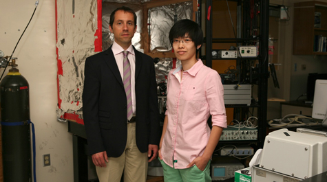 Zapping brain cells Christopher Del Negro, associate professor of applied science at William & Mary, and Ph.D. students Xueying "Snow" Wang and John Hayes (not pictured), have developed a method of identifying and killing individual brain cells in vitro. Photo by Stephen Salpukas
A team of William & Mary neuroscientists has developed a “search and destroy” method of identifying—then zapping—brain cells individually. It’s a laboratory technique that researchers expect to drive important advances in the understanding of how cumulative and progressive cell loss affects neural function.
The work has great implications for the increased understanding of a broad range of problems in neuroscience, including neurodegenerative diseases, explains Christopher Del Negro, associate professor of applied science at William & Mary. The team also includes graduate student Xueying “Snow” Wang and John Hayes, a former graduate student in applied science. The results were published in a paper titled “Cumulative lesioning of respiratory interneurons disrupts and precludes motor rhythms in vitro” in a recent issue of the Proceedings of the National Academy of Sciences.
The process can be used to study how brain cells devoted to various functions work together in networks, he explained. The William & Mary group focused on the neural respiratory control center known as the pre-Bötzinger Complex, although the technique could be valuable in the study of other neural motor functions such as locomotion or chewing, whose rhythms depend on the coordinated cell groups known as central pattern generator networks. Del Negro says his group’s work will provide greater understanding of the neurology of respiration, which could one day help clinicians who treat patients afflicted with neurodegenerative disorders.
“There are many neurological diseases. Some of the most famous are Alzheimer’s, Parkinson’s, amyotrophic lateral sclerosis, also known as Lou Gehrig’s disease. These diseases are known to be fatal,” Del Negro said. “However, people with Parkinson’s and Alzheimer’s rarely die of the disease they have. They actually die of respiratory failure.”
He explained that diseases such as Parkinson’s primarily attack neurons in the striatum, the upper-level brain. “But there’s collateral damage. Neurons are dying all over the central nervous system, including the respiratory part of the medulla, the lower brain stem,” he said. “When you have Parkinsonism in a patient, the things you recognize are motor deficits, not breathing deficits, but at some point, that person could stop breathing.”
The new in vitro technique allows researchers to chart the progressive loss of function in neural networks by identifying and then killing brain cells individually. Del Negro explains that the process mimics the progressive cell-by-cell loss characteristic of neurodegenerative diseases.
“This is a way to model diseases in a dish in the laboratory,” he said. “It’s a tool that can be used to model, mimic and potentially understand disease progression in the brain better.”
Del Negro points out that the “search and destroy” technique also is efficient in the lab, in that it uses the same instrument for both the detection and the destruction of the individual cells. He credits Hayes, now associated with France’s Centre National de la Recherche Scientifique, with the innovation.
“Our system uses a laser microscope to look at brain tissue and detect individual brain cells,” Del Negro explained. “Then it turns the microscope into a destructive device, and we go back and kill off the cells that it formerly detected. And while it does this, it monitors the functionality of the network.”
Their work, which was funded by the National Institutes of Health, shows the pre-Bötzinger Complex loses its rhythmic function necessary for respiration after the death of around 18 percent of its neurons. Del Negro cautions that these results are likely not directly applicable to other neural networks governing other brain functions, but he said that researchers investigating many different areas of the brain could use the technique.
“We are exporting this technique to other laboratories so that they can learn how we do this and do it themselves,” he said.
