


New confocal microscope will get plenty of use
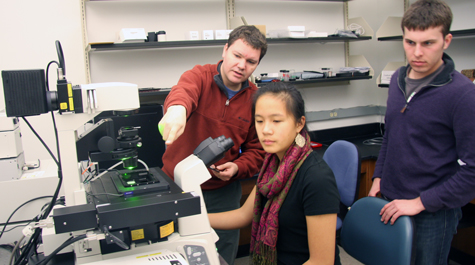 Increased capability Matthew Wawersik shows Andrea Lin '13 and Matthew Badgett '12 some of the features of the new confocal microscope installed in William & Mary's Integrated Science Center. The NSF-funded instrument will be used by a number of researchers and their students. Photo by Joseph McClain
Matthew Wawersik spends a lot of time looking at fruit flies. His lab uses these little bugs as a model to study germ line stem cell development. The addition of a state-of-the-art laser scanning confocal microscope allows Wawersik to see both his flies and his major research question from a fresh perspective.
This new laser scanning confocal microscope system was purchased via a Major Research Instrumentation (MRI) Grant through the National Science Foundation. Wawersik, associate professor of biology at William & Mary, was the lead principal investigator (PI) on the grant responsible for purchasing the new system.
“Because of this new system, the research that we can now do is on the level of the best and biggest medical schools in the country,” he said.
Co-PIs on the grant include Wawersik’s fellow biologists Lizabeth Allison, Margret Saha and Oliver Kerscher. “There are a lot of people throughout the department who are dreaming up ways to utilize the new system,” says Wawersik.
There are a variety of features on this new system that make it particularly exciting, including a resonance scanner, “basically just a fancy term for a really, really fast camera” explains Wawersik. The rapid shutter speed and high sensitivity of the resonance scanner facilitates live imaging, allowing Wawersik or another user to watch individual cells move three-dimensionally though tissue. The system is state of the art, and it can be upgraded as new technologies are developed and become available. Furthermore, Warwersik points out that the system is incredibly user-friendly.
“This system is more intuitive than the previous system that we had,” said Wawersik. “It’s incredibly nice in that respect, and with the right training you can just jump on. And it has additional very intuitive functions that you can do with minimal set-up; like live cell imaging over time.”
The new system is going to be an important tool in teaching in addition to its role in research. Wawersik plans on using the system for demonstrations in both upper-level biology and applied sciences courses. Furthermore, Wawersik stressed that the system will be accessible to students involved with research on campus.
“The students in our research labs are a major contributor to our success and our capacity to publish,” he said. “We’re already having our graduate students starting on it and our faculty is now learning to be comfortable with it. Once we have a reasonable lever of comfort and expertise, our undergraduates are going to be jumping on it and studying some questions that are critical to all of our labs.”
Scientists at larger institutions often have to pay for time with comparable confocal systems, but at William & Mary, time with the system will be free.
“It’s a departmental service by the four PIs. We obviously get a lot out of it because it’s critical to our research, but at other places you’d have to fork over the dollars for this service,” Wawersik explained. “Here we can have a system that is maintained by the department. It’s an incredible asset that doesn’t exist at most institutions around the country. It is rare and it really allows research to tick at William & Mary. It wouldn’t be feasible or accessible, if it weren’t for this case. The free nature allows us to say: ‘let’s do a microcopy demo on live imaging for a class.’ And, certainly, people are lining up to do it.”
