Biology Department microscopy resources include a laser scanning confocal microscope, automated and manual epifluorescent microscopes, fluorescent stereoscopes, a field emission scanning electron microscope (FESEM), and a transmission electron microscope (TEM).
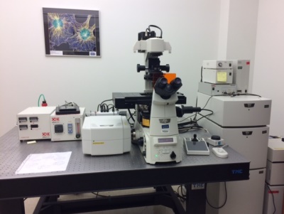
Nikon A1R inverted microscope equipped with resonant scanner, spectral imager, perfect focus technology and fully automated stage with temperate regulated growth chamber for live cell imaging. This confocal is equipped with 404, 488, 561, and 639 nm laser lines.
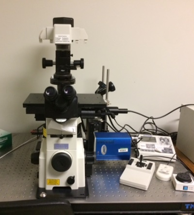
Fully automated Nikon TE2000 epifluorescent inverted microscope, equipped with a digital CCD camera
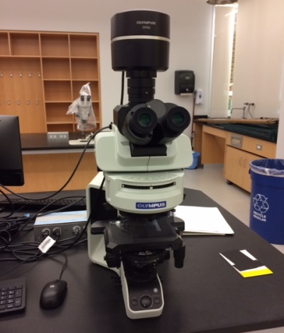
Manually operated Olympus BX53 epifluorescent microscope equipped with dual monochrome/color camera
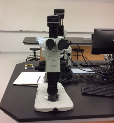
Olympus SZX16 fluorescent and brightfield stereoscope equipped with color camera
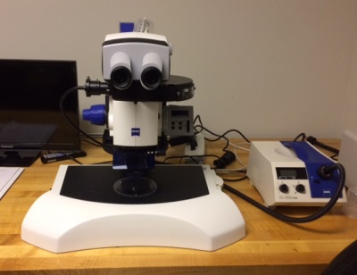
Zeiss Discovery.V12 fluorescent and brightfield stereoscope
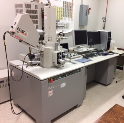
Hitachi S-4700 field emission scanning electron microscope (FESEM) equipped with EDX
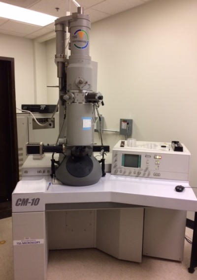
Philips FEI CM 10 transmission electron microscope typically run at 80 kV, capable of 100kV